





Most black spots on teeth are caused by tartar that builds up along the gumline, smoking, iron-containing medications, or pigment-producing bacteria in the mouth, and they can usually be cleaned away in a single clinic visit. However, not every dark-looking area is a stain: if there is darkening on a tooth's chewing surface or between two teeth, this is most often decay and will not go away with cleaning. A single tooth turning entirely gray or dark, on the other hand, is a different situation and usually points to an old injury.
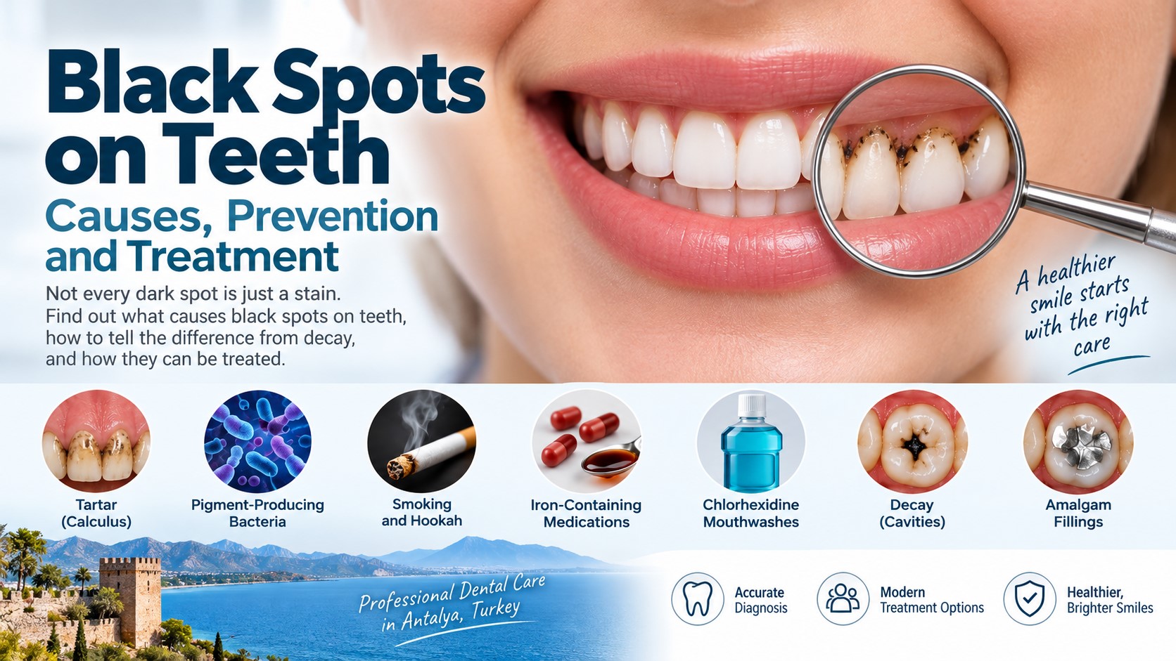
What Are the 7 Common Causes of Black Spots on Teeth?
- Tartar (calculus): Tartar that accumulates along and beneath the gumline darkens over time, often forming a near-black band on the back surface of the lower front teeth in particular.
- Pigment-producing (chromogenic) bacteria: Certain bacteria in the mouth combine with iron in saliva to leave a dotted or linear black band along the gumline. This is common in children and is discussed separately below.
- Smoking and hookah (waterpipe): Tar causes brownish discoloration at first, turning nearly black with long-term use; because it settles into microscopic cracks in the enamel surface, it does not come off with brushing.
- Iron-containing medications and syrups: Iron supplements used to treat anemia, especially in syrup form, cause temporary dark staining on the teeth.
- Chlorhexidine mouthwashes: Brown-black surface staining is a well-known side effect when used for longer than two weeks.
- Decay: Decay that has reached the dentin appears as a black cavity. Arrested (non-progressing) decay can also be hard and black.
- Amalgam fillings: Old silver-colored fillings can create a gray-black shadow in the adjacent tooth structure, and can also leave a permanent gray mark on the gum.
The black band seen in children
A thin black line made up of dots running along the gumline is a common finding in children that often worries parents. Its cause is not inadequate brushing; it is a compound formed by pigment-producing bacteria in the mouth combining with iron in saliva. For this reason, it is not directly related to sugar consumption either.
Children with this finding have sometimes been reported to have lower rates of decay, although the relationship is still being studied. In other words, the appearance is bothersome, but it is not itself a disease. It is removed in the clinic by polishing; however, because the bacterial flora remains the same, it can return within a few months. What is needed in this case is not harder brushing but regular check-ups and cleaning. For more on children's dental health, see the pediatric dentistry (pedodontics) page.
Black spot or decay? How to tell them apart
This distinction is the most important part of this page, because the two require completely different treatments.
A stain usually takes the form of a horizontal line along the gumline or discoloration spread across the surface; the tooth surface stays smooth and causes no sensitivity. Decay, on the other hand, is a localized darkening in the pits of the chewing surface or at the contact point between two teeth; you can feel roughness or a cavity on the surface, floss catches in that spot, and sensitivity to sweets or cold may accompany it.
A visual assessment alone is not always reliable; decay between teeth may not be visible during a routine visual examination and is often detected more reliably with bite-wing X-rays.
What does it mean when a single tooth darkens?
A single tooth that is noticeably darker than its neighbors, taking on a gray or black tone, is not a surface stain. Its most common cause is a past blow to the tooth: after trauma, the pulp (the tooth's nerve tissue) loses its vitality, and the blood byproducts released discolor the tooth from within. The injury may have happened years earlier.
In this case, no cleaning or whitening applied to the outer surface will make a difference. The tooth's vitality is assessed first; if needed, root canal treatment is performed, after which internal (non-vital) whitening applied inside the tooth can lighten the color. If the result is not sufficient, a crown becomes an option. The same appearance can also occur after an old root canal treatment.
How are black spots removed in the clinic?
- Scaling (tartar removal): Deposits along and beneath the gumline are removed with ultrasonic tips, usually resolving the discoloration completely.
- Air-polishing (air-powder polishing): This is the most effective method for stubborn surface stains caused by smoking and chromogenic bacteria.
- Filling: If the darkening is caused by decay, the decay is removed and a filling is placed.
- Root canal treatment and internal whitening: This is applied to a single tooth that has darkened due to trauma.
- Composite or crown: If the color cannot be lightened by any method, the tooth's surface is covered; see composite bonding.
Can it be removed at home?
Established black stains usually cannot be removed effectively with normal brushing at home and may require professional cleaning. Most of the methods people try at this point can actually cause harm: acidic mixtures such as lemon and baking soda may damage enamel, while abrasive charcoal products and aggressive brushing can contribute to enamel wear and gum recession. Enamel loss is irreversible, and it makes the darker dentin beneath the thinned enamel more visible.
At home, the main goal is to reduce the risk of new stains forming: brushing twice a day for two minutes each time, flossing daily, quitting smoking, and, if taking an iron supplement, using a straw and then rinsing the mouth afterward.
Preventing recurrence
Black stains tend to recur more often than yellow stains; those caused by chromogenic bacteria in particular can return a few months after cleaning. This does not mean the treatment failed. Check-ups and cleaning every six months, daily interdental cleaning, and quitting smoking significantly extend the interval before recurrence. If you use a chlorhexidine mouthwash, do not exceed the duration your dentist has specified.
Black Spot Treatment on Teeth in Antalya, Turkey
At our Hurma, Lara, and Güzeloba branches, the initial examination determines whether the darkening is a stain or decay; a bite-wing X-ray is evaluated for the areas between teeth. For surface stains, cleaning and polishing can be done on the same day. If a single tooth has darkened, a vitality test is performed first, and treatment is planned accordingly.
When Should You See a Dentist for a Black Spot on a Tooth?
With black discoloration, the real question isn't how to remove the stain, but whether it's actually a stain at all. Surface stains are cleaned in a single session; decay-related darkening, if left untreated, deepens over time and can progress all the way to needing root canal treatment. Since the two cannot always be told apart by eye, an examination should not be delayed when you notice a dark area that doesn't go away with brushing.
Frequently asked questions
Surface stains caused by tartar, smoking, and pigment-producing bacteria are usually removed in a single clinic session with scaling and air-polishing. If the darkening is caused by decay, it will not go away with cleaning and a filling is needed.
The most common causes are tartar, smoking, pigment-producing bacteria combining with iron in saliva, iron-containing medications and syrups, long-term use of chlorhexidine mouthwash, decay, and old amalgam fillings.
It results from a compound formed by pigment-producing bacteria in the mouth combining with iron in saliva; it is not directly related to inadequate brushing or sugar consumption. It is removed in the clinic by polishing, but it can return within a few months.
A stain usually appears as a horizontal line along the gumline, the surface stays smooth, and it causes no sensitivity. Decay, on the other hand, is a localized darkening on the chewing surface or between two teeth; roughness can be felt on the surface, and sensitivity may accompany it. Decay between teeth may not be visible during a routine visual examination and is often detected more reliably with bite-wing X-rays.
The most common cause is a past injury to the tooth, which may affect the pulp and cause discoloration from within. The tooth should first be assessed with a vitality test. If the pulp has lost vitality, root canal treatment may be required, followed by internal whitening where appropriate.
Established black stains usually cannot be removed effectively with normal brushing at home and may require professional cleaning. Acidic mixtures such as lemon and baking soda may damage enamel, while abrasive charcoal products and aggressive brushing can contribute to enamel wear and gum recession. All that can be done at home is to slow the formation of new stains.
It removes black discoloration caused by deposits along and beneath the gumline. For stubborn stains caused by smoking, air-polishing is applied in addition. Darkening caused by decay or trauma requires different treatment.
Those caused by pigment-producing bacteria in particular can return within a few months; this does not mean the treatment failed. Six-monthly check-ups, daily flossing, and quitting smoking extend the interval before recurrence.